Animal Eye Center of NJ’s ophthalmologist Dr. Brad Holmberg has some information to share about glaucoma and how you can protect your pet from vision loss.
Glaucoma is unfortunately a common disease in dogs that frequently results in permanent vision loss. Primary glaucoma, or that caused by a genetic or inherited component, is most common. Predisposed dog breeds include cocker spaniels, basset hounds, shiba inus, chow chows, shar peis, and arctic breeds. While any age can be affected, most dogs are affected in mid-life (around age 5-9 years old) with female dogs twice as likely to be affected as male dogs.
Early detection of glaucoma and early treatment are the best ways to prevent vision loss. Clinical signs of glaucoma can be obvious or subtle, but usually include redness of the white part of the eye (called episcleral congestion), a hazy or blue colored cornea (called corneal edema), a dilated pupil, and vision loss. As the signs are usually just in one eye, the change in vision may be difficult to detect. Some dogs appear quite painful and squint with more obvious signs, while others demonstrate their discomfort in a more subtle fashion, usually just being more lethargic.
Unlike humans with glaucoma (POAG) who have a slow elevation in intraocular pressure, dogs have a very rapid increase in pressure that causes damage immediately. Many times permanent vision loss can occur after only a few hours of elevated pressure. Therefore, glaucoma is a true emergency as every minute may make a difference in long-term visual outcome. Luckily, there are numerous medications and surgical procedures available for glaucoma and a veterinary ophthalmologist customizes the best treatment plan for each pet.
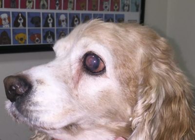
Meet Molly, a 7-year-old cocker spaniel that came to the Animal Eye Center of NJ for sudden redness and cloudiness of the left eye. As you can see, not only is the eye red and cloudy, but the pupil is dilated. Molly has acute glaucoma and her intraocular pressure was 38, with normal being between 10 and 20. Fortunately,
Molly still had some vision and aggressive medical therapy reduced her pressure to 11. She is currently managed on medical therapy alone, but we may pursue surgery in the future to better control her glaucoma long-term. The surgical procedure of choice is called cyclophotocoagulation with an anterior chamber shunt placement. This procedure uses a laser to selectively destroy sections of the “faucet” part of the eye to reduce inflow of fluid and then the shunt is placed to act as a new drain that increases the outflow of fluid.