Diabetes Mellitus and Ophthalmic Health: Key Considerations for the General Practitioner
News

Diabetes mellitus (DM) is a common endocrine disorder in dogs and cats with important ophthalmic implications, particularly in canine patients. Although systemic management is key to the long-term care of diabetic patients, ocular complications may develop rapidly, including diabetic cataracts, keratoconjunctivitis sicca (KCS), and diabetic retinopathy, which can significantly affect vision, comfort, and quality of life. Early recognition of these changes, appropriate ophthalmic monitoring, and timely referral to a veterinary ophthalmologist are important components of comprehensive diabetic patient management.
AEC patient Harley prior to cataract surgery (above), and after surgery (below).
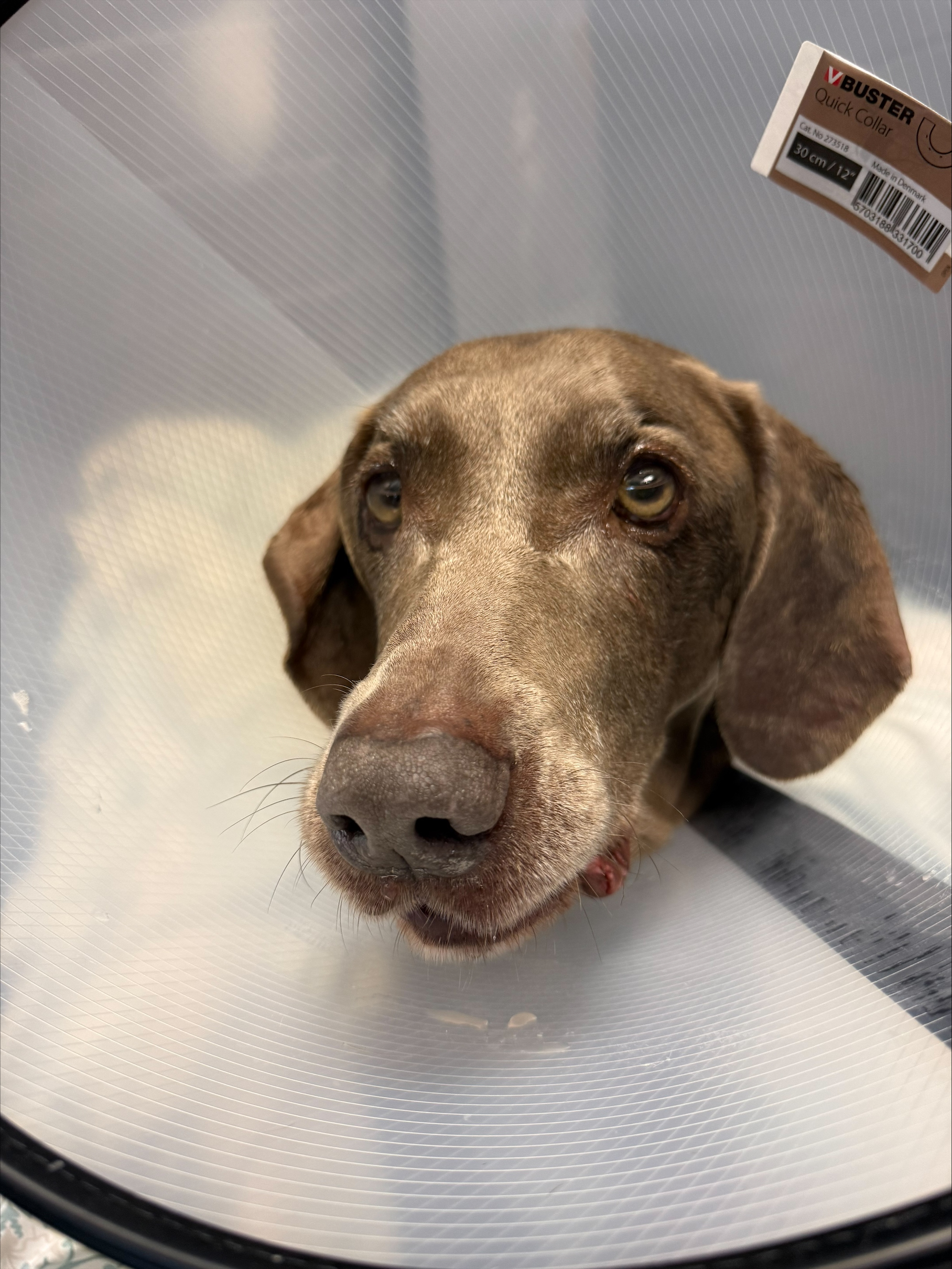
Diabetic Cataracts: Early Recognition and Timely Referral
Diabetic cataracts are among the most clinically significant ocular complications of DM in dogs and may develop and progress rapidly following diagnosis. One study reported that 80% of diabetic dogs developed cataracts within 16 months of diagnosis, with 60% developing cataracts within the first six months.¹ These findings underscore the importance of discussing the risk of cataract formation with owners at the time of DM diagnosis and incorporating ocular assessment into ongoing patient care.
Cataract formation in diabetic dogs is driven by increased activity of aldose reductase, an enzyme that converts excess glucose into sorbitol. Sorbitol accumulates within the lens, creating an osmotic gradient that promotes water influx and lens fiber swelling. This disrupts the orderly arrangement of lens fibers, compromises lens transparency, and ultimately results in cataract formation.²
Beyond vision impairment, diabetic cataracts may be associated with significant secondary complications, including lens-induced uveitis, glaucoma, and retinal detachment. These sequelae can result in ocular pain and irreversible vision loss. To prevent these complications, with cataract development in a DM canine patient, we recommend starting an NSAID topical solution once to twice daily and prompt referral to a veterinary ophthalmologist. Early evaluation allows for us to conduct pre-operative testing in a timely fashion, which includes a full ophthalmic exam as well as ocular ultrasound, gonioscopy, and electroretinogram, to assess the health and function of the eyes and to determine if a patient is a candidate for cataract surgery.
Keratoconjunctivitis Sicca and Diabetic Neuropathy
KCS, or dry eye disease, is another potential ocular complication seen in diabetic patients. Normal corneal sensory innervation is vital to normal tear production and protective blinking reflexes, both of which are essential for maintaining ocular surface health. Diabetic neuropathy may affect peripheral sensory and autonomic nerve function, potentially compromising corneal sensitivity, lacrimal gland function, and ocular surface protection¹.
Chronic hyperglycemia is suspected to contribute to diabetic nerve injury through metabolic and microvascular changes. For general practitioners, assessment of tear production and the ocular surface may be warranted when diabetic patients present with ocular irritation, conjunctival hyperemia, mucoid discharge, corneal changes, or reduced corneal sensitivity. Patients with suspected KCS should undergo appropriate diagnostic testing, including a Schirmer tear test, and receive treatment and follow-up based on the severity of disease.
Diabetic Retinopathy
Diabetic retinopathy is a recognized complication of DM in dogs, although it appears to be less prevalent and generally less severe than the disease observed in humans. In people, progressive retinal microvascular damage can lead to substantial visual impairment. In dogs, reported retinal changes include retinal hemorrhages and other vascular abnormalities, which do not necessarily progress to clinically significant vision loss¹.
Nevertheless, retinal abnormalities in diabetic patients warrant appropriate investigation rather than being presumed incidental or benign. Fundic examination, when feasible, can help identify posterior segment changes. Referral for a comprehensive ophthalmic examination is appropriate when retinal lesions are identified, visual deficits are suspected, or the fundus cannot be adequately evaluated.
Integrating Ophthalmic Care into Diabetic Patient Management
Ophthalmic assessment is an important consideration in the long-term management of diabetic patients, particularly dogs at risk for rapidly progressive cataracts. At diagnosis, general practitioners should counsel owners regarding the potential for cataract development, assess the eyes as part of the physical examination, and refer quickly for ophthalmic referral when abnormalities are detected.
Timely referral is especially important for newly identified or rapidly progressing cataracts, suspected lens-induced uveitis, elevated intraocular pressure, unexplained visual deficits, and concerning retinal changes. Collaboration between the primary care veterinarian and veterinary ophthalmologist supports early identification of ocular disease, appropriate intervention, and coordinated management of systemic and ophthalmic health.
References:
Miller, E. J., & Brines, C. M. (2018). Canine diabetes mellitus associated ocular disease.
Topics in Companion Animal Medicine
, 33*(1)*, 29–34.
Gelatt, K. N., Ben-Shlomo, G., Gilger, B. C., Hendrix, D. V. H., Kern, T. J., & Plummer, C. E. (Eds.). (2021).
Veterinary Ophthalmology
(6th ed., Vols. 1–2). Wiley-Blackwell.